The Dissertations Sticky

For the discussion of catfish systematics. Post here to draw our attention to new publications or to discuss existing works.
Post Reply
User avatar
bekateen
Posts: 9254
Joined: 09 Sep 2014, 17:50
I've donated: $40.00!
My articles: 4
My images: 141
My cats species list: 144 (i:102, k:37)
My aquaria list: 36 (i:13)
My BLogs: 44 (i:149, p:2680)
My Wishlist: 35
Spotted: 182
Location 1: USA, California, Stockton
Location 2: USA, California, Stockton
Contact:

Evolutionary Ecology of Loricariid Catfishes

Post by bekateen »

MODERATOR'S NOTE: This paper was published on 2 November 2022: viewtopic.php?t=51252

Black, C. (2022). Evolutionary Ecology of Loricariid Catfishes. Ph.D. dissertation, Auburn University.

https://etd.auburn.edu/handle/10415/8312

PDF: https://etd.auburn.edu/bitstream/handle ... sAllowed=y
Abstract
How extrinsic (like functional constrains and ecological interactions) and intrinsic (like modularity and integration) interactions drive diversification is a formative area of evolutionary biology. In this dissertation, I explore the phenotypic diversification of the armored catfishes using geometric morphometrics, stable isotope analyses, and phylogenetic comparative methods. I found that the armored catfish body is highly modularized, with varying degrees of integration between each module, suggesting that interactions within and between modules influence morphological evolution. Additionally, slight changes in modularity and integration patterns in clades may have allowed for diversification along a specific trajectory. When focused on the oral jaw shape, I found that traditional and automated processes captured shape more effectively when all jaw components were combined. Although ecological traits do not play a role in jaw shape, there was a correlation between clades with diverse diets and fast evolutionary rates of shape. These results suggest that shape is not constrained to diet and that similarly shaped jaws coupled with different types of teeth could allow the fishes to feed on a wide range of materials. Finally, I built a vector-based analysis, baseline‐standardized isotopic vector analysis (BaSIVA) to visualize dietary variation while accounting for isotopic discrepancies between locations. Results from BaSIVA delineate trophic groups better than traditional trophic positioning methods while accounting for variation in basal resources, suggesting BaSIVA should be the standard for vector‐based stable isotope analysis in riverine environments with similar baseline resources.
Image
Find me on YouTube & Facebook: http://youtube.com/user/Bekateen1; https://www.facebook.com/Bekateen
Buying caves from https://plecocaves.com? Plecocaves sponsor Bekateen's Fishroom. Use coupon code bekateen for 15% off your order.
Also, for you Swifties: Https://youtu.be/ZUKdhXL3NCw
User avatar
Jools
Expert
Posts: 16107
Joined: 30 Dec 2002, 15:25
My articles: 198
My images: 948
My catfish: 237
My cats species list: 87 (i:237, k:1)
My BLogs: 7 (i:10, p:202)
My Wishlist: 23
Spotted: 450
Location 1: Middle Earth,
Location 2: Scotland
Interests: All things aquatic, Sci-Fi, photography and travel. Oh, and beer.
Contact:

Re: Diversity of Trichomycterus in the Rio Doce basin

Post by Jools »

bekateen wrote: 17 Oct 2018, 05:37 Ten new species proposed:
  • sp. nov.
  • T. barrocus sp. nov.
  • T. brucutu sp. nov.
  • T. illuvies sp. nov.
  • T. melanopygius sp. nov.
  • T. ipatinguensis sp. nov.
  • T. pussilipygius sp. nov.
  • T. sordislutum sp. nov.
  • T. vinnulus sp. nov.
  • T. tantalus sp. nov.
This one now published. What do I do - create a new post?

Jools
User avatar
bekateen
Posts: 9254
Joined: 09 Sep 2014, 17:50
I've donated: $40.00!
My articles: 4
My images: 141
My cats species list: 144 (i:102, k:37)
My aquaria list: 36 (i:13)
My BLogs: 44 (i:149, p:2680)
My Wishlist: 35
Spotted: 182
Location 1: USA, California, Stockton
Location 2: USA, California, Stockton
Contact:

Re: Diversity of Trichomycterus in the Rio Doce basin

Post by bekateen »

Jools wrote: 20 Nov 2022, 12:15 [
This one now published. What do I do - create a new post?

Jools
That's what I've been doing, then I link the two posts together with a moderators note. Perfect example is my thesis post immediately above yours here. Take a look:
bekateen wrote: 01 Aug 2022, 14:44 MODERATOR'S NOTE: This paper was published on 2 November 2022: viewtopic.php?t=51252

Black, C. (2022). Evolutionary Ecology of Loricariid Catfishes. Ph.D. dissertation, Auburn University.

https://etd.auburn.edu/handle/10415/8312
Image
Find me on YouTube & Facebook: http://youtube.com/user/Bekateen1; https://www.facebook.com/Bekateen
Buying caves from https://plecocaves.com? Plecocaves sponsor Bekateen's Fishroom. Use coupon code bekateen for 15% off your order.
Also, for you Swifties: Https://youtu.be/ZUKdhXL3NCw
User avatar
Jools
Expert
Posts: 16107
Joined: 30 Dec 2002, 15:25
My articles: 198
My images: 948
My catfish: 237
My cats species list: 87 (i:237, k:1)
My BLogs: 7 (i:10, p:202)
My Wishlist: 23
Spotted: 450
Location 1: Middle Earth,
Location 2: Scotland
Interests: All things aquatic, Sci-Fi, photography and travel. Oh, and beer.
Contact:

Re: The Dissertations Sticky

Post by Jools »

OK, I will do, I didn't see that as I was at the original post a bit back up the thread. All should be done now,

Cheers,

Jools
User avatar
bekateen
Posts: 9254
Joined: 09 Sep 2014, 17:50
I've donated: $40.00!
My articles: 4
My images: 141
My cats species list: 144 (i:102, k:37)
My aquaria list: 36 (i:13)
My BLogs: 44 (i:149, p:2680)
My Wishlist: 35
Spotted: 182
Location 1: USA, California, Stockton
Location 2: USA, California, Stockton
Contact:

Two new species of Pariolius; New genus on the horizon

Post by bekateen »

new species
new species
Coming soon (maybe): new genus
may get a new generic designation, but is not yet renamed.

Fuster, D. R. F. (2022). Análise filogenética de Heptapteridae gill, 1861 e taxonomia integrativa de Heptapterus bleeker, 1858 (Heptapterinae: heptapterini). Ph.D. Dissertation. Graduate Program in Animal Biology, Instituto de Biociências da Universidade Federal do Rio Grande do Sul. Porto Alegre, Brazil.
https://www.lume.ufrgs.br/handle/10183/245860
PDF: https://www.lume.ufrgs.br/bitstream/han ... sequence=1
ABSTRACT
The Heptapteridae family contains 23 genera and 231 valid species that are specific in a wide range of freshwater habitats from southern Mexico to northern Argentina. A current phylogenetic systematics of Heptapteridae is significantly shaped by an unpublished morphology-based analysis of most extant genera carried out over 20 years ago. We provide a new multilocus molecular phylogenetic hypothesis encompassing 19 of 23 valid genera in Heptapteridae, including all valid genera of Brachyglaniini (4 of 4), 11 of 14 valid genera of Heptapterini and 66% of all valid species of Heptapterini (58 of 88, plus many new species). The analysis includes 15 type species from the 23 valid genera of Heptapteridae. The present work is based on a multilocus analysis of five molecular markers, being three mitochondrial markers (COI, cytochrome oxidase subunit I; Cyt b: cytochrome be ND2: NADH dehydrogenase subunit 2) and two nuclear markers (RAG2: recombination activating 2, and Glyt: glycosyltransferase), and have recovered generally well-resolved and consistently supported phylogenies. Based on these results, we provide a new suprageneric classification within Heptapteridae, subdivided into: Heptapterinae (comprising Brachyglaniini and Heptapterini) and Rhamdiinae (containing Rhamdiini and Goeldiellini); five new subtribes were recovered within Heptapterini. This work includes an integrative analysis of the genus , type genus of the family, that is redefined and limited to four valid species. It includes the description of a new genus sister of Heptapterus and containing two new species. Finally, we present the description of new species within Heptapteridae.
RESUMO
A família Heptapteridae contém 23 gêneros e 231 espécies válidas que são encontradas em uma ampla gama de habitats de água doce do sul do México ao norte da Argentina. A sistemática filogenética atual de Heptapteridae é significativamente moldada por uma análise baseada em morfologia da maioria dos gêneros existentes, realizada há mais de 20 anos e não publicada. Nós fornecemos uma nova hipótese filogenética molecular abrangendo 19 dos 23 gêneros válidos em Heptapteridae, incluindo todos os gêneros válidos de Brachyglaniini (4 de 4), 11 dos 14 gêneros válidos de Heptapterini e 66% de todas as espécies válidas de Heptapterini (58 de 88; além de muitas espécies não descritas). A amostragem inclui 15 espécies tipo dos 23 gêneros válidos de Heptapteridae. O presente trabalho, baseado em uma análise multilocus de cinco marcadores moleculares, 3 marcadores mitocondriais (COI, cytochrome oxidase subunit I; Cyt b: cytochrome b e ND2: NADH dehydrogenase subunit 2), e dois marcadores nuclear (RAG2: recombination activating 2, e Glyt: glycosyltransferase), produziu filogenias geralmente consistentes, bem resolvidas e fortemente suportadas. Com base nesses resultados, fornecemos uma nova classificação supragenérica dentro de Heptapteridae subdividida em: Heptapterinae (contendo Brachyglaniini e Heptapterini) e Rhamdiinae (contendo Rhamdiini e Goeldiellini); dentro de Heptapterini foram reconhecidas cinco subtribos novas. O trabalho inclui uma análise integrativa do gênero espécie tipo da família onde o gênero foi redefinido e limitado a 4 quatro espécies validas, com a descrição de um gênero novo irmão de Heptapterus contendo duas espécies novas. Finalmente apresentamos descrições de novas espécies de Heptapteridae.
Image
Find me on YouTube & Facebook: http://youtube.com/user/Bekateen1; https://www.facebook.com/Bekateen
Buying caves from https://plecocaves.com? Plecocaves sponsor Bekateen's Fishroom. Use coupon code bekateen for 15% off your order.
Also, for you Swifties: Https://youtu.be/ZUKdhXL3NCw
User avatar
bekateen
Posts: 9254
Joined: 09 Sep 2014, 17:50
I've donated: $40.00!
My articles: 4
My images: 141
My cats species list: 144 (i:102, k:37)
My aquaria list: 36 (i:13)
My BLogs: 44 (i:149, p:2680)
My Wishlist: 35
Spotted: 182
Location 1: USA, California, Stockton
Location 2: USA, California, Stockton
Contact:

Systematics of the genus Rhyacoglanis

Post by bekateen »

Rodrigues, Jefferson Luan Crispim. (2023). Systematics of the genus Rhyacoglanis Shibatta & Vari, 2017 (Siluriformes: Pseudopimelodidae). Paulista State University (Unesp). Available at: < http://hdl.handle.net/11449/239252 >.

https://repositorio.unesp.br/handle/11449/239252
Abstract
Within the great diversity of Neotropical fish, we can highlight the Pseudopimelodidae family, a group of catfish widely distributed throughout South America, whose representatives have been characterized by the presence of a yellow body color with dark bars, which associates them with the popular name of bumblebee. catfishes, covering two subfamilies, namely, Batrochoglaninae and Pseudopimelodinae, which includes the genus . In order to improve understanding and understanding, this Course Completion Work is divided into two chapters: In the first, the molecular delimitation of the species of the genus Rhyacoglanis was carried out using the cytochrome c oxid markerase subunit I (COI) to carry out a delimitation analysis of species of the genus. Fourteen samples of muscle tissue from species of the genus Rhyacoglanis, from the hydrographic basins of the Paraná, Paraguay, Xingu, Tapajós and Madeira rivers, covering a wide geographic distribution for the genus, were used. For the species delimitation analysis, we used the ASAP program, which, in its first partition, indicated the existence of four independent lineages and in its second partition it indicated five lineages, namely, R. annulatus, R. pulcher, R. variolosus and two new species, one for the Tapajós river basin and another for the Xingu river basin. In the second chapter, a new species of Rhyacoglanis is described for the rio Jamanxim, rio Tapajós basin.
Image
Find me on YouTube & Facebook: http://youtube.com/user/Bekateen1; https://www.facebook.com/Bekateen
Buying caves from https://plecocaves.com? Plecocaves sponsor Bekateen's Fishroom. Use coupon code bekateen for 15% off your order.
Also, for you Swifties: Https://youtu.be/ZUKdhXL3NCw
User avatar
bekateen
Posts: 9254
Joined: 09 Sep 2014, 17:50
I've donated: $40.00!
My articles: 4
My images: 141
My cats species list: 144 (i:102, k:37)
My aquaria list: 36 (i:13)
My BLogs: 44 (i:149, p:2680)
My Wishlist: 35
Spotted: 182
Location 1: USA, California, Stockton
Location 2: USA, California, Stockton
Contact:

Re: The Dissertations Sticky

Post by bekateen »

Obafunmiso, H. (2023). Behavioral response to conspecific sounds in Loricariid catfishes (Pterygoplichtys pardalis and Otocinclus vittatus). Master's thesis, 3661, Biology Department, Western Kentucky University. Online Aug 2023.

https://digitalcommons.wku.edu/theses/3661/
Abstract
Sound production is a means of communication among many fish species. In fishes, sound is produced through various mechanisms, but in the family Loricariidae, known as armored suckermouth catfishes, sound is produced primarily through pectoral fin spine stridulation. Previous experiments have described the sounds produced and shown the mechanism of sound production in two species of loricariid catfishes, and , but the functional significance of loricariid sounds is still unknown. To address this question, I examined the behavioral responses of Pterygoplichthys pardalis and Otocinclus vittatus to conspecific calls. Individual fish (N=10 for P. pardalis) or groups of 20 fish (N=4 groups for O. vittatus) were acclimated to an aquarium for at least 24 h. Individual P. pardalis were video recorded for 2 minutes with no sound, plus another 2 minutes with a playback of either a 500 Hz tone control or conspecific call through an underwater speaker. In contrast, O. vittatus was video recorded for 5 minutes with no sound, with an additional 5 minutes of either conspecific call or 500 Hz tone stimuli. This procedure was repeated for each individual or group using either a 500 Hz tone and conspecific call for playback so that each individual or group received both stimuli. I hypothesized that P. pardalis would avoid, while O. vittatus would be attracted to, the conspecific sound-emitting speaker, respectively. The rationale for this hypothesis is that P. pardalis produces calls when it is under duress, so it may be an alarm call, while O. vittatus produces calls spontaneously in large groups of fish, suggesting it may be a cohesion call or involved in other intraspecific interactions. P. pardalis showed an increased activity level to conspecific sound compared to the 500 Hz tone, although movement in general was minimal, while O. vittatus exhibited a short-lived response to conspecific calls by moving toward the speaker with the sound source. Since both species showed a minimal behavioral change to sound playbacks, more research is needed to better understand the function of sound production in loricariid catfishes.
Image
Find me on YouTube & Facebook: http://youtube.com/user/Bekateen1; https://www.facebook.com/Bekateen
Buying caves from https://plecocaves.com? Plecocaves sponsor Bekateen's Fishroom. Use coupon code bekateen for 15% off your order.
Also, for you Swifties: Https://youtu.be/ZUKdhXL3NCw
User avatar
bekateen
Posts: 9254
Joined: 09 Sep 2014, 17:50
I've donated: $40.00!
My articles: 4
My images: 141
My cats species list: 144 (i:102, k:37)
My aquaria list: 36 (i:13)
My BLogs: 44 (i:149, p:2680)
My Wishlist: 35
Spotted: 182
Location 1: USA, California, Stockton
Location 2: USA, California, Stockton
Contact:

Reproductive biology of Loricariichthys anus

Post by bekateen »

Selle, D.C. (2023). Characterization of the reproductive biology of fiddle (Loricariichthys anus). Master's Dissertation in Zootechnics – Animal Production, Faculty of Agronomy, Federal University of Rio Grande do Sul, Porto Alegre, RS, Brazil. (69 p.) March, 2023. Creative Commons license by-nc-sa 2.5

Selle, D.C. (2023). Caracterização da biologia reprodutiva de violinha (Loricariichthys anus). Dissertação de Mestrado em Zootecnia – Produção Animal, Faculdade de Agronomia, Universidade Federal do Rio Grande do Sul, Porto Alegre, RS, Brasil. (69 p.) março, 2023. Creative Commons license by-nc-sa 2.5

https://lume.ufrgs.br/handle/10183/263176
PDF: https://lume.ufrgs.br/bitstream/handle/ ... sAllowed=y
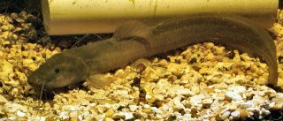
ABSTRACT - The "violinha" (
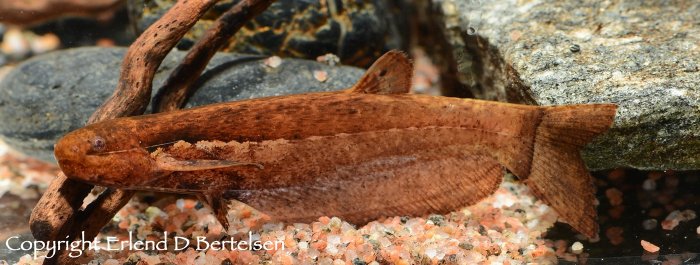
) has been commercially prominent in southern Brazil in the last three decades, becoming a vital fishery resource. Given its high market acceptance in Rio Grande do Sul, it is the second most consumed native fish in the Holy Week in 2021, being seen as a species with great potential for aquaculture. To perform the sexual characterization of the species and elucidate critical biological aspects for the development of captive breeding protocols, the present study was carried out the following analyses aiming to characterize the annual reproductive cycle: sexual dimorphism, gonadosomatic index (GSI), gonadal histology of males and females, sperm kinetics, sperm concentration, and sperm morphology. To analyze the variations of these factors throughout the year, six males and six females were collected monthly in the Guaíba River from November 2021 to October 2022, totaling 144 animals (51.02 ± 16.61 g and 22.3 ± 1.96 cm). The animals were anesthetized, euthanized, measured, and weighed, and their gonads were obtained by dissection and weighed on a precision scale. Part of each male gonad was macerated to get semen (for kinetic, concentration, and morphological analysis), and the other part, along with the female gonads, was fixed in Glutaraldehyde 2,5% for further histological analysis. The morphological evaluation of the animals was based on evaluation studies of the Loricariidae family. Was obtained a single characteristic for dimorphism the lower lip of males (7.8 ± 2.71 mm), which presented elongation of 2 to 3 times its standard size (5.3 ± 0.70 mm), besides showing darkening on the sides and final portion of the lower lip, during the months of November to March. As for the IGS analysis, the data were observed, month by month for males and females, presenting the following variation, respectively: 0.41 ± 0.10 and 4.73 ± 0.51 (November); 0.35 ± 0.11 and 5.53 ± 1.33 (December); 0.41 ± 0.11 and 4.29 ± 1.91 (January); 0.55 ± 0.09 and 3.29 ± 3.57 (February); 0.16 ± 0.09 and 0.68 ± 0.94 (March); 0.17 ± 0.07 and 0.90 ± 0.40 (April); 0.06 ± 0.03 and 0.69 ± 0.59 (May); 0.24 ± 0.05 and 0.94 ± 0.42 (June); 0.18 ± 0.12 and 0.72 ± 0.28 (July); 0.28 ± 0.21 and 0.73 ± 0.59 (August); 0.14 ± 0.03 and 1.53 ± 1.40 (September); 0.21 ± 0.13 and 2.79 ± 3.93 (October). The fixed gonads were embedded in paraplast and sectioned on a manual microtome to assemble slides, stained with hematoxylin and eosin, and analyzed by light microscopy to identify the phases of gonadal maturation over the months. For females, the analyzes indicate that between November and March, they presented mature oocytes, in addition to demonstrating that there is more than one occurrence of total spawning during the reproductive period. On the other hand, males did not present a defined reproductive period, with the occurrence of spermatozoa throughout the year, but with low volumes, and from December on, the testicles are already observed regressing, with a large volume of spermatogonial cells. For the sperm kinetics analysis (Motility - MOT; curvilinear velocity - VCL; straight line velocity - VSL; mean displacement velocity - VAP and progressivity - PROG), performed with the CASA software, the semen samples were diluted in Hank's Solution and later activated with distilled water. There were differences (p>0.05) for MOT between the analyzed periods of November, January, and March (25.58 ± 0.05; 1.97 ± 0.02 and 2.11 ± 0.005 %); VCL (58.8 ± 4.47; 37.59 ± 5.29 and 41.28 ± 6.9 μm/s, respectively) and VAP (40.1 ± 4.99; 20.71 ± 3.04 and 22.87 ± 5.61 μm/s). For sperm concentration, the samples were placed in a Neubauer chamber where a 5-field subjective count was performed. Subsequently, the value found was converted, presenting an average of 9,741,600 ± 1,208,834 spermatozoa/ml of semen, but only during the reproductive period (November to March). In the other months, we did not obtain enough semen volume to perform the analysis. For morphology, 200 spermatozoa were counted in each analysis, getting the following values: normal sperm 103 ± 21.79; loose head 69 ± 14.77; short tail 7 ± 3.68; degenerated head 6 ± 4.16; macrocephaly 4 ± 3.16; microcephaly 2 ± 2.09. With the data obtained from IGS and gonadal histology, it can be stated that the reproductive period of L. anus occurs between November and February in the Guaíba River, corroborating the information that the species presents sexual dimorphism only during the reproductive period. It is also possible to state that the species shows a low amount of viable spermatozoa during the reproductive period, besides a low sperm concentration, when compared to other species of the order Siluriforme or even the family Loricariidae, and this may be a bottleneck for the reproduction of the species in captivity.
Keywords: Sperm kinetics; sperm concentration; sexual dimorphism; IGS; sperm morphology.
RESUMO - A violinha (Loricariichthys anus) vem se destacando comercialmente no sul do Brasil nas últimas três décadas, tornando-se um importante recurso pesqueiro. Visto sua elevada aceitação de mercado no Rio Grande do Sul, é o segundo peixe nativo mais consumido na semana santa em 2021, sendo vista como uma espécie com grande potencial para aquicultura. Com intuito de realizar a caracterização sexual da espécie e elucidar aspectos biológicos importantes para o desenvolvimento de protocolos de reprodução em cativeiro, o presente estudo realizou as seguintes análises com o objetivo de caracterizar o ciclo reprodutivo anual: Dimorfismo sexual; Índice Gonadossomático (IGS); Histologia gonadal de machos e fêmeas; Cinética espermática; Concentração espermática; e Morfologia espermática. Para analisar as variações desses fatores ao longo do ano, foram coletados no rio Guaíba, de novembro de 2021 a outubro de 2022, seis machos e seis fêmeas ao mês, totalizando 144 animais (51,02 ± 16,61 g e 22,3 ± 1,96 cm). Os animais foram anestesiados, eutanasiados, mensurados e pesados e suas gônadas foram obtidas por dissecação, pesadas em balança de precisão. Parte de cada uma das gônadas masculinas foram maceradas afim de se obter sêmen (para as analises cinéticas, de concentração e morfológica), e a outra parte, juntamente com as gônadas femininas, fixadas em glutaraldeído 2,5% para posterior análise histológica. A avaliação morfológica dos animais baseou-se em trabalhos de avalição para Família Loricariidae, obtendo-se um único ponto de dimorfismo, o lábio inferior dos machos (7,8 ± 2,71 mm), que apresentou alongamento de 2 a 3 vezes o seu tamanho normal (5,3 ± 0,70 mm), além de apresentar escurecimento nas laterais e porção final do lábio inferior, durante os meses de novembro a março. Quanto à análise do IGS, os dados foram observados, mês a mês para machos e fêmeas foram, apresentação a seguinte variação, respectivamente: 0,41 ± 0,10 e 4,73 ± 0,51 (novembro); 0,35 ± 0,11 e 5,53 ± 1,33 (dezembro); 0,41 ± 0,11 e 4,29 ± 1,91 (janeiro); 0,55 ± 0,09 e 3,29 ± 3,57 (fevereiro); 0,16 ± 0,09 e 0,68 ± 0,94 (março); 0,17 ± 0,07 e 0,90 ± 0,40 (abril); 0,06 ± 0,03 e 0,69 ± 0,59 (maio); 0,24 ± 0,05 e 0,94 ± 0,42 (junho); 0,18 ± 0,12 e 0,72 ± 0,28 (julho); 0,28 ± 0,21 e 0,73 ± 0,59 (agosto); 0,14 ± 0,03 e 1,53 ± 1,40 (setembro); 0,21 ± 0,13 e 2,79 ± 3,93 (outubro). As gônadas fixadas foram incluídas em paraplast e seccionadas em micrótomo manual para a montagens das lâminas, as quais foram coradas em hematoxilina e eosina e analisadas por microscopia óptica para identificação das fases de maturação gonadal ao longo dos meses. Para fêmeas as análises indicam que entre novembro e março apresentaram oócitos maduros, além de demonstrar que há mais de uma ocorrência de desova total durante o período reprodutivo. Já os machos não apresentaram período reprodutivo definido, com ocorrência de espermatozoides durante todo o ano, porém em baixos volumes e, a partir de dezembro, já se observa os testículos regredindo, com um grande volume de espermatogônias. Para a análise da cinética espermática (Motilidade - MOT; velocidade curvilínear - VCL; velocidade em linha reta - VSL; velocidade média de deslocamento - VAP e progressividade - PROG), realizada com o software CASA, as amostras de sêmen foram diluídas em Solução de Hank's e posteriormente ativadas com água destilada. Houve diferença (p>0,05) para MOT entre os períodos analisados de novembro, janeiro e março (25,58 ± 0,05; 1,97 ± 0,02 e 2,11 ± 0,005 %); VCL (58,8 ± 4,47; 37,59 ± 5,29 e 41,28 ± 6,9 μm/s, respectivamente) e VAP (40,1 ± 4,99; 20,71 ± 3,04 e 22,87 ± 5,61 μm/s). Para concentração espermática, as amostras foram posicionadas em câmara de Neubauer onde realizou-se a contagem de subjetiva de 5 campos e, posteriormente, o valor encontrado foi convertido, apresentando a média de 9.741.600 ± 1.208.834 espermatozoides/ml de sêmen, mas apenas durante o período reprodutivo (novembro a março). Nos demais meses não se obteve volume suficiente de sêmen para realizar a análise. Para morfologia foram contabilizados 200 espermatozoides em cada análise, obtendo-se os seguintes valores: Espermatozoides normais 103 ± 21,79; Cabeça solta 69 ± 14,77; Cauda curta 7 ± 3,68; Cabeça degenerada 6 ± 4,16; Macrocefalia 4 ± 3,16; Microcefalia 2 ± 2,09. Com os dados obtidos de IGS e histologia gonadal, pode-se afirmar que o período reprodutivo de L. anus ocorre entre os meses de novembro e fevereiro no rio Guaíba, corroborando com a informação de que a espécie apresenta dimorfismo sexual apenas durante o período reprodutivo. Também é possível afirmar que a espécie apresenta uma baixa quantidade de espermatozoides viáveis durante o período reprodutivo, além de baixa concentração espermática, quando comparadas à outras espécies da ordem Siluriforme ou, até mesmo, da família Loricariidae, podendo este ponto ser um gargalo para reprodução da espécie em cativeiro.
Palavras-chave: Cinética espermática; concentração espermática; dimorfismo sexual; IGS; morfologia espermática.
Attachments
Figure 1. Illustrative image of a specimen of the yellow-bellied catfish Loricariichthys anus. Photo: Douglas Cosme Selle (Collection carried out in October 2022).
Figure 9. Images exemplifying the three groups used to distinguish the color of the upper and lower lips of the viola (Loricariichthys anus): A) specimen with Normal lip; B) specimen with dark edges of the lower and upper lips; and C) specimen with dark Perimeter of the lower and upper lips.
Figure 3. Image of a male specimen of yellow-bellied catfish (Loricariichthys anus) incubating fertilized eggs on its lower lip. Photo: Daniela Pereira da Rosa.
Figure 6. Image demonstrating the embryonic development of Loricaria coximensis. Photo: IMASUL, 2019.
Figure 4. Larval development of the yellow-bellied catfish (Loricariichthys anus). Photo: Daniela Pereira da Rosa.
Image
Find me on YouTube & Facebook: http://youtube.com/user/Bekateen1; https://www.facebook.com/Bekateen
Buying caves from https://plecocaves.com? Plecocaves sponsor Bekateen's Fishroom. Use coupon code bekateen for 15% off your order.
Also, for you Swifties: Https://youtu.be/ZUKdhXL3NCw
User avatar
bekateen
Posts: 9254
Joined: 09 Sep 2014, 17:50
I've donated: $40.00!
My articles: 4
My images: 141
My cats species list: 144 (i:102, k:37)
My aquaria list: 36 (i:13)
My BLogs: 44 (i:149, p:2680)
My Wishlist: 35
Spotted: 182
Location 1: USA, California, Stockton
Location 2: USA, California, Stockton
Contact:

Ontogeny and phylogeny in Trichomycteridae

Post by bekateen »

Reis, V. J. C. (2023). Ontogeny and phylogeny in Trichomycteridae (Teleostei, Siluriformes): patterns in the development of morphological complexes. Doctoral Thesis, Museu de Zoologia, University of São Paulo, São Paulo. doi:10.11606/T.38.2023.tde-17082023-144452. Retrieved 2023-09-23, from www.teses.usp.br
https://doi.org/10.11606/T.38.2023.tde-17082023-144452
https://www.teses.usp.br/teses/disponiv ... 452/en.php
Abstract
This thesis offers the first large-scale study on the ontogeny of the musculoskeletal system in Trichomycteridae and its implications for understanding the phylogenetic relationships and evolution of the family. Trichomycteridae is a monophyletic group of neotropical catfish with exceptionally rich morphological diversity. This diversity is reflected in a wide range of habitats and trophic niches. The feeding habits of the group vary from invertivores to strict hematophagy, the latter a unique case in Actinopterygii. Using developmental series and juvenile specimens representing most trichomycterid lineages, detailed descriptions of the development of various musculoskeletal systems of the head, such as the mandibular complex, opercular apparatus, branchial skeleton, suspensorium, and neurocranium are presented. Such information, new for the most part, provides a solid framework for formulating and testing hypotheses of homology, some of which representing long-standing controversies in Siluriformes, such as the supraoccipital, parietal, pterotic, extrascapular, and posttemporal. Observations also permit direct mapping of transformation sequences and connections between highly divergent conditions in adult specimens, such as the mouth apparatus of juveniles and adults of Vandelliinae. Results obtained are incorporated into new morphological and total evidence phylogenetic analyses with broader taxonomic and data density than previously available, allowing a detailed understanding of the diversification history of Trichomycteridae. Those results are combined with developmental information to provide an ontogenetic framework for testing a new method for detecting heterochronies designed to map paedo- and peromorphic phenomena in the family. The most relevant phylogenetic results are that Microcambevinae probably result from long-branched attraction, and the identification of a new clade composed of Stenolicmus ix and Ammoglanis pulex, previously assigned to Sarcoglanidinae, and the removal of Potamoglanis from Tridentinae. The synergism of phylogenetic hypothesis with ontogeny reveals wide-evolutionary patterns, showing that Trichomycteridae is a mostly paedomorphic family, a ground plan upon which numerous internested peramorphic apomorphies have evolved. One major event of miniaturization occurred at the base of a large clade, here named the miniature clade. Within that clade, there are various events of further reduction in size. Surprisingly, within the miniature clade there is also an event of reverse size-increase, a phenomenon detected not only by phylogenetic structure but also by marks left in the structure and development of different anatomical complexes.
Image
Find me on YouTube & Facebook: http://youtube.com/user/Bekateen1; https://www.facebook.com/Bekateen
Buying caves from https://plecocaves.com? Plecocaves sponsor Bekateen's Fishroom. Use coupon code bekateen for 15% off your order.
Also, for you Swifties: Https://youtu.be/ZUKdhXL3NCw
User avatar
bekateen
Posts: 9254
Joined: 09 Sep 2014, 17:50
I've donated: $40.00!
My articles: 4
My images: 141
My cats species list: 144 (i:102, k:37)
My aquaria list: 36 (i:13)
My BLogs: 44 (i:149, p:2680)
My Wishlist: 35
Spotted: 182
Location 1: USA, California, Stockton
Location 2: USA, California, Stockton
Contact:

Evolutionary biogeography of catfishes

Post by bekateen »

Janzen, F. (2023). Evolutionary biogeography of catfishes (Siluriformes, Actinopterygii): The influence of habitat and landscape on gene flow and genetic diversification. PhD Dissertation, Ottawa-Carleton Institute of Biology, University of Ottawa, Canada.
https://ruor.uottawa.ca/handle/10393/45512
Abstract
A fundamental goal of evolutionary biology is to understand what processes have led to the great diversity of organisms we see today. An important factor of diversification is an organism’s environment. Abiotic factors can shape the evolutionary trajectory of species by affecting fundamental mechanisms of evolution, including mutation, gene flow, genetic drift, and natural selection. In my thesis, I investigated how abiotic factors, such as habitat and landscape, have influenced the genetic diversification of catfishes (Siluriformes). More specifically, I compared genetic data within and between species to understand how natural barriers have shaped the origins and evolutionary trajectory of species. In Chapter 1, I investigated whether habitat preferences and segregation of breeding populations in lacustrine-like and fluvial habitats affected the genetic structure of a sympatric population of channel catfish (). In Chapter 2, I elucidated the origins of cave species within North American catfishes (Ictaluridae), determining whether they shared a common ancestor or evolved in parallel from surface-dwelling ancestors. In Chapter 3, I tested whether impermeable and semi-permeable boundaries between South American river basins have restricted gene flow and resulted in potentially new species within the widespread ornate pim catfish (). In Chapter 4, I determined whether orogenesis and river capture corresponded with speciation events and cladogenesis within Neotropical long-whiskered catfishes (Pimelodidae). Throughout my thesis, I observed evolutionary patterns related to gene flow, vicariance, and dispersal. Physical barriers imposed on populations often coincided with genetic diversification and allopatric speciation. These barriers reduced gene flow, allowing populations to genetically diverge in response to unique selective pressures. As these barriers changed over time, dispersal opportunities may have further promoted diversification as species radiated into new areas. I also observed that ecological gradients, such as water chemistry, may have facilitated parapatric speciation; however, differences between habitats do not always restrict gene flow. Given that patterns of genetic diversification and speciation are not uniform across the tree of life, it is important for evolutionary biologists to document trends among different taxa to elucidate macroevolutionary patterns.
Image
Find me on YouTube & Facebook: http://youtube.com/user/Bekateen1; https://www.facebook.com/Bekateen
Buying caves from https://plecocaves.com? Plecocaves sponsor Bekateen's Fishroom. Use coupon code bekateen for 15% off your order.
Also, for you Swifties: Https://youtu.be/ZUKdhXL3NCw
User avatar
bekateen
Posts: 9254
Joined: 09 Sep 2014, 17:50
I've donated: $40.00!
My articles: 4
My images: 141
My cats species list: 144 (i:102, k:37)
My aquaria list: 36 (i:13)
My BLogs: 44 (i:149, p:2680)
My Wishlist: 35
Spotted: 182
Location 1: USA, California, Stockton
Location 2: USA, California, Stockton
Contact:

Mitochondrial haplotype variations of Clarias camerunensis and C. gariepinus in Cameroon

Post by bekateen »

Piyumi Sandaruwani De Alwis. 2023. Mitochondrial haplotype variations of Clarias camerunensis and C. gariepinus in Cameroon. Master's Thesis, Department of Marine Biology, Pukyong National University

https://repository.pknu.ac.kr:8443/hand ... .oak/33281
https://repository.pknu.ac.kr:8443/bits ... meroon.pdf
ABSTRACT

The air-breathing walking catfish (Clariidae: Clarias) consists of 32 species that are native to African freshwater environments. Because of their complicated taxonomy and variability, species-level identification of this group is difficult. Prior to this study, biological and ecological investigations were limited to a single species, Clarias gariepinus, resulting in a skewed understanding of genetic diversity in African waters. The 63 mitochondrial Cytochrome c oxidase subunit 1 (COI) gene sequences of Clarias camerunensis and Clarias gariepinus from Cameroon's Nyong River were produced here. Both C. camerunensis and C. gariepinus species preserved appropriate intra-species (2.7% and 2.31%) and inter-species (6.9% to 16.8% and 11.4% to 15.1%) genetic distances with other Clarias congeners located in African and Asian/Southeast Asian drainages. The mtCOI sequences indicated 13 and 20 distinct haplotypes of C. camerunensis and C. gariepinus, respectively. TCS networks indicated unique haplotypes of C. camerunensis and shared haplotypes of C. gariepinus in African waters. The various species delimitation methodologies (ABGD and PTP) identified a total of 20 and 22 molecular operational taxonomic units (MOTUs), respectively. Among the two Clarias species studied, we discovered more than one MOTU in C. camerunensis, which is compatible with population structure and tree topology results. The phylogeny created by Bayesian Inference analysis convincingly distinguished C. camerunensis and C. gariepinus from other Clarias species, with strong posterior probability supports. This mitogenome of C. camerunensis was circular (16,511 bp long) and contained 13 protein-coding genes (PCGs), two ribosomal RNAs (rRNAs), 22 transfer RNAs (tRNAs), and a single AT-rich regulatory region. The heavy strand contains 28 genes, whereas the light strand contains ND6 and eight tRNA genes. The mitochondrial genome of C. camerunensis is AT-biased (56.89%), as shown in other Clarias species. The comparative analysis found that the majority of Clarias species had six overlapping and 11 intergenic spacer regions. The ATG start and TAA end codons were used to launch and terminate the majority of PCGs. Except for tRNA-serine, the tRNAs of C. camerunensis folded into the characteristic cloverleaf secondary structure. The positioning of the conserved domains in the control area was consistent in all Clarias species with substantially varied nucleotides in conservation blocks I. Maximum-likelihood and Bayesian-based matrilineal phylogenies clearly split all Clarias species into five clades based on their known ranges (South China, Sundaland, Indochina, India, and Africa). The TimeTree study found that the two major clades (Indo-Africa and Asia) of Clarias species may have separated during the Paleogene (28.66 MYA). The current study investigates the presence of putative cryptic diversity and allopatric speciation of C. camerunensis in African drainages. Furthermore, the current study reveals that C. gariepinus has reduced genetic diversity across its native and imported range, which may have been caused by improper aquaculture operations. The study suggests applying a similar strategy to the same and related species from multiple river basins to reveal the full variety of Clarias species in Africa and other nations. Our findings show that Indian species (Clarias dussumieri) and African species (C. camerunensis and C. gariepinus) split during the Paleogene, while South Chinese species (Clarias fuscus) and Sundaland species (Clarias batrachus) split from Indochinese species (Clarias macrocephalus) split during the Neogene through independent colonization. This biotic interaction pattern emphasizes the importance of topography and geological events in dictating the evolutionary history of Clarias species. The enrichment of mitogenomic data and various nuclear loci from their original range or type place will prove Clarias species' real diversity in African and Asian nations.
Image
Find me on YouTube & Facebook: http://youtube.com/user/Bekateen1; https://www.facebook.com/Bekateen
Buying caves from https://plecocaves.com? Plecocaves sponsor Bekateen's Fishroom. Use coupon code bekateen for 15% off your order.
Also, for you Swifties: Https://youtu.be/ZUKdhXL3NCw
User avatar
bekateen
Posts: 9254
Joined: 09 Sep 2014, 17:50
I've donated: $40.00!
My articles: 4
My images: 141
My cats species list: 144 (i:102, k:37)
My aquaria list: 36 (i:13)
My BLogs: 44 (i:149, p:2680)
My Wishlist: 35
Spotted: 182
Location 1: USA, California, Stockton
Location 2: USA, California, Stockton
Contact:

Loricariidae from the Miocene, State of Acre, Brazil

Post by bekateen »

Jacó, Tiago Ricardo Fernandes. 2023. Loricariidae (Ostariophysi: Siluriformes) from the Miocene (Solimões Formation, Acre Basin), State of Acre, Brazil. Dissertation (Master's) - State University of Rio Grande do Sul, Secretariat of the Environment and Infrastructure of Rio Grande do Sul, Academic Master's Degree in Systematics and Conservation of Biological Diversity, Unit in Porto Alegre, 2023. 94 pp, il.
https://repositorio.uergs.edu.br/xmlui/ ... 56789/2989
https://repositorio.uergs.edu.br/xmlui/ ... sAllowed=y
ABSTRACT
Today, Loricariidae (Siluriformes) is the largest catfish family in the Neotropical region, with 1,039 species considered valid. The taxa are easily recognized by their morphological characteristics: elongated body, bony plates, odontodes, compressed head and suckermouth and for having specialized feeding habits. Some loricariids such as Hypostomus group cochliodon, Panaque and Panaqolus, maintain a diet based preferentially on wood. This dietary preference is related to the spoon-shaped dentition observed in these taxa. The present study aims to record fossil teeth of Loricariidae from an outcrop located on the right bank of the Juruá River, named PRJ- 26, Municipality of Marechal Thaumaturgo, State of Acre (AC), Brazil. The specimens were obtained from conglomeratic layer sediments of the Solimões Formation (Neogene) by the screen washing method, and after sorting under a stereoscopic microscope they were deposited in the Scientific Collection of the Paleontology Laboratory of the Campus Floresta of Universidade Federal do Acre, Cruzeiro do Sul, AC. This material is composed of 28 dental fragments, most of which present fractures and loss of the implantation stems. The teeth are assigned to two subfamilies of Loricariidae: Hypostominae and Loricariinae. The Hypostominae, which hold most specimens, were classified as: (i) Hypostomus cochliodon group (11 specimens), presenting the dental crowns wide, of various sizes, and the presence of a lateral cuspid, with different shapes and angles; (ii) cf. Panaque sp. (11 specimens) with a strong, concave and unicuspid crown, (iii) and cf. Ancistrus sp. (one specimen) with a simple, unicuspid, long, spatulate crown with a rounded apex. The Loricariinae, in turn, were identified at the tribe level: (i) Loricariini indet. (Four specimens), with bifid crowns, two partially fused lobes, one lobe being prominent, and with a rounded apex; (ii) Hartiini indet., a single specimen showing a bifid crown, its lobes being fused and delimited only by a groove. Knowledge of loricariid fossils in the South American Cenozoic includes records for the Miocene of Argentina, Colombia, and Peru and Venezuela, most of which are classified as Loricariidae indet. Fossil studies of outcrop PRJ-26 are still in early stages, so it is not safe to conclusively state an age for the outcrop. However, similarities are noted between the conglomerate channel of outcrop PRJ-26 with other localities that include records of Loricariidae remainsin the South American Cenozoic, especially in the northern region of South America, which correspond to middle-superior Miocene ages.
KEY WORDS: Western Amazon, Cascudos, Teeth, Fossils, Morphology.
Image
Find me on YouTube & Facebook: http://youtube.com/user/Bekateen1; https://www.facebook.com/Bekateen
Buying caves from https://plecocaves.com? Plecocaves sponsor Bekateen's Fishroom. Use coupon code bekateen for 15% off your order.
Also, for you Swifties: Https://youtu.be/ZUKdhXL3NCw
User avatar
bekateen
Posts: 9254
Joined: 09 Sep 2014, 17:50
I've donated: $40.00!
My articles: 4
My images: 141
My cats species list: 144 (i:102, k:37)
My aquaria list: 36 (i:13)
My BLogs: 44 (i:149, p:2680)
My Wishlist: 35
Spotted: 182
Location 1: USA, California, Stockton
Location 2: USA, California, Stockton
Contact:

Comparative anatomy and taxonomic revision of the genus Pareiodon

Post by bekateen »

Victório, O.G. (2024). Comparative anatomy and taxonomic revision of the genus Pareiodon Kner, 1855 (Siluriformes: Trichomycteridae), a scavenger fish from the Amazon basin. Doctoral dissertation, Universidade de São Paulo. DOI https://doi.org/10.11606/D.59.2024.tde-10072024-104826
Abstract in English
The family Trichomycteridae is one of the main representatives of neotropical Siluriformes and presents wide morphological and ecological diversity, with members displaying remarkable specializations in their way of life and feeding habits. The subfamily Stegophilinae contains individuals with elongated bodies and a feeding habit typically based on mucus and scales. , a member of Stegophilinae, is a monotypic genus represented by Pareiodon microps Kner, 1855. The species has a wide distribution in the Amazon basin and stands out from other members of the subfamily in terms of its feeding habits. P. microps has a diet based on pieces of other dying or dead fish, and this ecological dynamic is associated with several characteristics of the oral region that were the focus of our observations. Considering the wide distribution, monotypy, and distinct feeding habit of the species, our project aimed to investigate the anatomy of the group to contribute to the understanding of its taxonomy and evolutionary history. External morphology, osteology, facial musculature, and the lateral line system were chosen for our investigations. The choice of the facial musculature complex was based on the association of this elements with foraging, respiration, and food capture activities. Comparing the muscle elements with other representatives of Trichomycteridae revealed the existence of characteristics exclusively present in Pareiodon microps and potentially associated with the evolution of carrion feeder habits. The lateral line system was found to be highly conserved among the analyzed representatives of Trichomycteridae, but the number of grooves present in the trunk's medial line showed potential taxonomic value. Additionally, a discussion is raised about the presence of superficial neuromasts on the ventral surface of the trunk in some Trichomycteridae members, a condition not previously reported in the literature. Despite the wide distribution, morphological characteristics are highly conserved among individuals from all basins where P. microps occurs. Thus, the monotypy of P. microps is reinforced by our observations. Diagnoses previously attributed to the group were revisited, with the presence of an internal process on the dentary being the only one questioned. Additionally, three new diagnoses for the group are proposed, all related to facial musculature and the lateral line system. An extensive anatomical description of the species was provided, with emphasis on osteology and facial musculature, accompanied by discussions about the evolution of carrion-feeder habits. The descriptions and discussions aim to contribute to the understanding of the species and the evolution of feeding habits in Trichomycteridae.
Keywords: Evolution, Myology, Neotropical fauna, Stegophilinae, Taxonomy

Victório, O.G. (2024). Anatomia comparada e revisão taxonômica do gênero Pareiodon Kner, 1855 (Siluriformes: Trichomycteridae), um peixe carniceiro da bacia Amazônica (Doctoral dissertation, Universidade de São Paulo). DOI https://doi.org/10.11606/D.59.2024.tde-10072024-104826
Abstract in Portuguese
A família Trichomycteridae é uma das principais representantes de Siluriformes neotropicais e apresenta ampla diversidade morfológica e ecológica, com membros que possuem marcantes especializações em seu modo de vida e alimentação. A subfamília Stegophilinae contém indivíduos de corpo alongado e hábito alimentar tipicamente baseado em muco e escamas. Pareiodon, membro de Stegophilinae, é um gênero considerado monotípico, sendo representado por Pareiodon microps Kner, 1855. A espécie é tida como amplamente distribuída na bacia amazônica e se apresenta como uma exceção em relação aos demais membros da subfamília no que diz respeito ao hábito alimentar. Pareiodon microps possui dieta baseada em pedaços arrancados de outros peixes moribundos ou mortos, sendo que essa dinâmica ecológica está associada a diversas características da região oral que foram alvo de nossas observações. Tendo em vista a ampla distribuição, a monotipia e o distinto hábito alimentar da espécie, nos propusemos a investigar a anatomia do grupo a fim de contribuir com a compreensão de sua taxonomia e história evolutiva. A morfologia externa, osteologia, musculatura facial e o sistema da linha lateral foram escolhidos como alvo de nossas investigações. A escolha pelo complexo da musculatura facial se baseou na associação desses elementos com as atividades de forrageamento, respiração e captura de alimento. A comparação dos elementos da musculatura com outros representantes de Trichomycteridae revelou a existência de características exclusivamente presentes em Pareiodon microps e, adicionalmente, nossas observações sobre o comportamento da espécie indicam que tais características podem estar associadas à evolução do hábito carniceiro. O sistema da linha lateral se mostrou bastante conservado entre os representantes de Trichomycteridae analisados, mas a quantidade de sulcos presentes na linha medial do tronco apresentou potencial valor taxonômico. Adicionalmente, é levantada uma discussão acerca da presença de neuromastos superficiais na superfície ventral do tronco de alguns representantes de Trichomycteridae, condição não reportada previamente na literatura. Apesar da ampla distribuição, as características morfológicas são muito conservadas entre os indivíduos das bacias hidrográficas nas quais P. microps ocorre. Desta forma, a monotipia do gênero Pareiodon é confirmada pelas nossas análises. As diagnoses previamente atribuídas ao grupo foram revisitadas, sendo que a presença de um processo interno no dentário foi a única questionada. Adicionalmente, são propostas três novas diagnoses para Pareiodon microps, todas fundamentadas em singularidades da musculatura facial e do sistema da linha lateral. Foi fornecida uma extensa descrição anatômica da espécie, com ênfase na osteologia e musculatura facial, acompanhada de discussões acerca da evolução do hábito carniceiro. As descrições e discussões realizadas buscam colaborar com a compreensão da espécie e da evolução do hábito carniceiro em Trichomycteridae.
Image
Find me on YouTube & Facebook: http://youtube.com/user/Bekateen1; https://www.facebook.com/Bekateen
Buying caves from https://plecocaves.com? Plecocaves sponsor Bekateen's Fishroom. Use coupon code bekateen for 15% off your order.
Also, for you Swifties: Https://youtu.be/ZUKdhXL3NCw
User avatar
bekateen
Posts: 9254
Joined: 09 Sep 2014, 17:50
I've donated: $40.00!
My articles: 4
My images: 141
My cats species list: 144 (i:102, k:37)
My aquaria list: 36 (i:13)
My BLogs: 44 (i:149, p:2680)
My Wishlist: 35
Spotted: 182
Location 1: USA, California, Stockton
Location 2: USA, California, Stockton
Contact:

Comparative morphology of lateral line canals across catfish families

Post by bekateen »

Almeida, M.A. (2024). Morphology, development and diversity of canals of the laterosensory system in Siluriformes (Actinopterygii; Ostariophysi). (Doctoral dissertation, University of São Paulo).

Almeida, M.A. (2024). Morfologia, desenvolvimento e diversidade dos canais do sistema latero-sensorial em Siluriformes (Actinopterygii; Ostariophysi) (Doctoral dissertation, Universidade de São Paulo).

https://doi.org/10.11606/T.59.2024.tde-10072024-144552
https://www.teses.usp.br/teses/disponiv ... 552/en.php
Abstract
The laterosensory system or lateral-line system is present in fishes and aquatic amphibians, playing a central role in behaviors such as predator evasion, prey capture and school organization. The functional units in this mechanoreceptor system are the neuromasts, which can be distributed throughout the head and trunk, inside canals (canal neuromasts) and on the skin surface (superficial neuromasts). The canals of the lateral-line system have proven to be a valuable source of characters for taxonomic and phylogenetic studies. However, features of this sensory system still largely unknown for several groups of fishes, including families in the Siluriformes order, popularly known as "catfishes". The order is among one of the most diverse groups in the superorder Ostariophysi. In this study, a comparative analysis was carried out in order to describe patterns of the canals of the laterosensory system among representatives of the families of the order Siluriformes. Data from observations on specimens of 40 valid families, prepared using different techniques, were combined with information available in the literature, regarding: canal identification, numbers of canal segments, connections between canals and canal-bone association. Tubules and multiple pores of the lateral-line canals were analyzed in an ontogenetic series (76 specimens from 22.7 - 90.6 mm SL) of , Pimelodidae family, with the aim of observing the change from simple to multiple tubules and pores and comparing with the conditions observed in adults of other catfish families. Among the members of the Siluriformes order, the following canals were identified: supraorbital, infraorbital, otic, preoperculomandibular, postotic, temporal and trunk. A rostral portion of the infraorbital canal or ethmoid commissure, as well as a supratemporal canal or commissure, were not observed. Relevant variations were observed in the number of canal segments in the supraorbital, infraorbital, preoperculomandibular and trunk canals, with substantial reductions and losses found in catfishes families from Loricarioidei lineage, 'Big Asia' clade and (Aspredinidae). Detailed observation about connections between canals, added to information on the development of P. corruscans, allowed a better interpretation of the union between the supraorbital, infraorbital and otic canals, as well as between the otic, postotic and preoperculomandibular canals. However, the main variations in canals connections were observed in (Cetopsidae). There is a very preserved canal-bone association, with small variations in the tubular-shaped ossifications nasal, infraorbitals, supra and subpreopercule and the presence of the extrascapular bone. The multiple tubules and pores in the cephalic lateral-line canals in P. corruscans arose in a certain size range during development. The condition of multiple pores appears several times among representatives of the Siluroidei lineage. A difference in the degree of these multiple pores was noted, between representatives of the same family and between different families. The results presented in this study indicate a great potential for the features about lateral-line canals to better understand the diversity in the Siluriformes order and their evolutionary relationships, with some of them deserving more in-depth studies.
Keywords: Catfishes, Lateral-line, Neuromasts
Resumen
O sistema látero-sensorial ou sistema da linha lateral está presente em peixes e anfíbios aquáticos, participando de comportamentos como, evasão de predadores, captura de presas e organização de cardumes. As unidades funcionais que constituem este sistema mecanorrecptor são os neuromastos, que podem estar distribuídos pela cabeça e tronco dos organismos, dentro de canais (neuromastos de canais) ou na superfície da pele (neuromastos superficiais). Os canais da linha lateral têm demonstrado ser uma valiosa fonte de caracteres em estudos taxonômicos e filogenéticos. No entanto, características desse sistema ainda são pouco ou completamente desconhecidas para vários grupos de peixes, incluindo famílias da ordem Siluriformes, peixes conhecidos popularmente como "bagres". Essa ordem está entre um dos agrupamentos mais diversos da superordem Ostariophysi. Neste estudo, uma análise comparativa foi realizada afim de descrever padrões dos canais do sistema látero-sensorial entre os representantes das famílias da ordem Siluriformes. Os dados provenientes das observações em espécimes de 40 famílias válidas, preparados com diferentes técnicas, foram combinados com as informações disponíveis na literatura sobre: identificação dos canais, número de segmentos, conexões e associação com ossificações. Túbulos e poros múltiplos dos canais da linha lateral foram analisados em uma série ontogenética (76 espécimes entre 22,7 - 90,6 mm CP) de , família Pimelodidae, com o objetivo de observar a mudança de tubos e poros simples para múltiplos nessa espécie e comparar com as condições observadas encontradas em outras famílias de bagres. Entre os membros da ordem Siluriformes foram identificados os canais: supraorbital, infraorbital, ótico, pré-opérculo-mandibular, pós-ótico, temporal e tronco. Não foi observada uma porção rostral do canal infraorbital ou comissura etmoidal, bem como um canal ou comissura supratemporal. Foram encontradas variações consideráveis no número de segmentos de canal nos canais supraorbital, infraorbital, pré-opérculo-mandibular e tronco, sendo as maiores reduções e perdas em bagres das famílias agrupadas na linhagem Loricarioidei, no clado 'Big Asia' e em (Aspredinidae). A observação detalhada das conexões entre os canais, somada às informações do desenvolvimento de P. corruscans, permitiram interpretar melhor a união entre os canais supraorbital, infraorbital e ótico, bem como entre os canais ótico, pós-ótico e pré-opérculo-mandibular. A presença da conexão interorbital variou consideravelmente entre táxons de diferentes famílias. No entanto, as maiores variações em conexões dos canais foram observadas em (Cetopsidae). Há uma associação canal-ossificações muito preservada, com pequenas variações nas ossificações tubulares nasal, infraorbitais, supra- e sub-pré-opérculo e a presença de um osso extra-escapular. Os múltiplos túbulos e poros nos canais cefálicos da linha lateral em P. corruscans surgiram em uma determinada faixa de tamanho durante o desenvolvimento. A condição de múltiplos poros surge diversas vezes entre os representantes da linhagem Siluroidei. Foi notada uma diferença no grau desses múltiplos poros, tanto entre representantes de uma mesma família, quanto entre diferentes famílias. Os resultados apresentados neste estudo indicam um grande potencial das características dos canais da linha lateral para compreender melhor a diversidade da ordem Siluriformes e suas relações de parentesco evolutivo, com algumas delas merecendo estudos mais aprofundados.
Palavras-chave: Bagres, Linha lateral, Neuromastos
Image
Find me on YouTube & Facebook: http://youtube.com/user/Bekateen1; https://www.facebook.com/Bekateen
Buying caves from https://plecocaves.com? Plecocaves sponsor Bekateen's Fishroom. Use coupon code bekateen for 15% off your order.
Also, for you Swifties: Https://youtu.be/ZUKdhXL3NCw
User avatar
bekateen
Posts: 9254
Joined: 09 Sep 2014, 17:50
I've donated: $40.00!
My articles: 4
My images: 141
My cats species list: 144 (i:102, k:37)
My aquaria list: 36 (i:13)
My BLogs: 44 (i:149, p:2680)
My Wishlist: 35
Spotted: 182
Location 1: USA, California, Stockton
Location 2: USA, California, Stockton
Contact:

Morphological development and osteological larvae and juvenile of lophiosilurus alexandri

Post by bekateen »

Assega, F.M., 2024. Desenvolvimento morfológico e osteológico de larvas e juvenis de Lophiosilurus alexandri Steindachner, 1877 (Siluriformes: Pseudopimelodidae), da Bacia do Rio São Francisco.

https://repositorio.uel.br/handle/123456789/14048
Abstract: This dissertation was divided into three chapters. The first deals with the general introduction that will briefly address the characteristics of Siluriformes and some useful tools for the taxonomy of specimens in the larval and juvenile periods. The second chapter aimed to follow the morphological development of the pacamã, Lophiosilurus alexandri, from the larval period to the juvenile period, through anatomical descriptions, and to observe the growth mode of the larval and juvenile stages, throughout its skeletal development, through analytical regression models. The third chapter dealt with describing the skeletal development of L. alexandri and, for this, specimens in the larval and juvenile periods were diaphanized and dissected, to observe the development mode of their structures. In this study, the skull, paired and odd fins were analyzed.
Abstract
Resumo: Esta Dissertação foi dividida em três capítulos, o primeiro trata da introdução geral que vai abordar brevemente as características dos Siluriformes e algumas ferramentas úteis para a taxonomia de exemplares nos períodos larvais e juvenil O segundo capítulo teve como objetivo acompanhar o desenvolvimento morfológico do pacamã, Lophiosilurus alexandri, desde o período larval até o período juvenil, por meio de descrições anatômicas, e observar o modo de crescimento dos estágios larvais e juvenil, ao longo do seu desenvolvimento esquelético, por meio de modelos analíticos de regressão O terceiro capítulo tratou de descrever o desenvolvimento esquelético do L alexandri e, para isso, foram diafanizados e dissecados exemplares nos períodos larvais e juvenil, para observar o modo de desenvolvimento de suas estruturas, nesse estudo foram analisados o crânio, as nadadeiras pares e ímpares
Keywords
Bagre (Peixe), Morfologia, Ontogenia, Peixe, Crescimento, Catfishes, Morphology, Ontogeny, Fish - Growth
Image
Find me on YouTube & Facebook: http://youtube.com/user/Bekateen1; https://www.facebook.com/Bekateen
Buying caves from https://plecocaves.com? Plecocaves sponsor Bekateen's Fishroom. Use coupon code bekateen for 15% off your order.
Also, for you Swifties: Https://youtu.be/ZUKdhXL3NCw
Post Reply

Return to “Taxonomy & Science News”